Internal medicine Jefferson Northeast Hospital Philadelphia, Pennsylvania, United States
Clinical Scenario or Case: A 63-year-old female with a past medical history of hypertension, type 2 diabetes mellitus, and a known left renal mass under surveillance was found to have an incidentally discovered right atrial mass on transthoracic echocardiogram. The patient was asymptomatic at the time of evaluation.
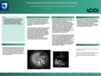
A contrast-enhanced CT scan revealed a large left renal mass with tumor extension into the inferior vena cava (IVC). Subsequent abdominal MRI demonstrated a centrally located 5 cm left renal mass with tumor thrombus extending cephalad through the IVC into the right atrium, consistent with a Level IV thrombus. The patient underwent open left radical nephrectomy, retroperitoneal lymph node dissection (RPLND), IVC thrombectomy, and sternotomy with cardiopulmonary bypass. Final pathology was consistent with clear cell renal cell carcinoma.
Evidence/Literature Review: Renal cell carcinoma (RCC) accounts for approximately 3% of all cancers, with over 80,000 new cases and nearly 15,000 deaths estimated in 2023. Tumor thrombus (TT) occurs in 4–10% of RCC cases, most commonly involving the renal vein (10–18%) and less frequently the inferior vena cava (IVC) (4–23%). 1% of cases, the thrombus can extend into the right atrium. Clear cell carcinoma is the predominant histologic subtype in RCC cases associated with TT.
Unique Aspects of Case: The RA thrombus was unexpectedly discovered on echocardiography—a rare initial clue for diagnosing RCC. Despite the presence of a Level IV (RA-involving) tumor thrombus, the patient was clinically stable and asymptomatic, which is uncommon. Initially considered to be a cardiac mass versus thrombus, the final diagnosis required advanced imaging and multidisciplinary input.
Complex surgical approach involving both urology and cardiothoracic teams.
Recommendations/Conclusions: RCC with tumor thrombus to the RA is a rare presentation with significant clinical challenges. An uncommon and often clinically silent presentation that underscores the importance of vigilant imaging and multidisciplinary management.